Author(s): Maalej Yosra*, Kharrat Malek, Zinelabidine Karim and Zbiba Walid
Cerulean or blue-dot cataracts are early onset bilateral cataracts with concentric layers of bluish-white opacities.
Visual acuity is usually well preserved and rarely necessitates cataract extraction before adult life.
Cerulean cataracts are a rare form of cataract characterized by blue and white opacifications scattered in the nucleus and cortex of the lens. Progression of cerulean cataracts is slow and often result in posterior subcapsular opacification that requires lens extraction in adults. Patients with cerulean cataract do not have any associated systemic comorbidities.
A 30-year-old woman presented with a gradual decrease in vision in the left eye for many years. The best-corrected visual acuity was 10/10 and 1/10 in the right and left eye, respectively. Examination of both eyes releveled multiple blue and white colored opacities spread throughout the cortex of lens more prominent and radially oriented in the center of the left eye. We note the presence of posterior subcapsular cataract in the right eye (Figure 1).
The intraocular pressure and the posterior segment examination were normal in both eyes. Cerulean cataracts are hereditary ocular disease (autosomal dominant) characterized by the presence of concentric layers of bluish-white opacities in the mid-periphery [1].
Appearing at the outer edge of the foetal lens nucleus, these opacities may involve the adult nucleus and the cortex in the form of concentric circles [2].
Because newborns are often asymptomatic until 18-24 months of age Cerulean cataracts are considered a form of developmental cataract rather than a true congenital cataract [2].
The progression of this rare type of cataract is slow; it can progress to a posterior subcapsular cataract.
Cerulean cataract may not become visually significant until the third to fourth decades when lens extraction may be helpful.
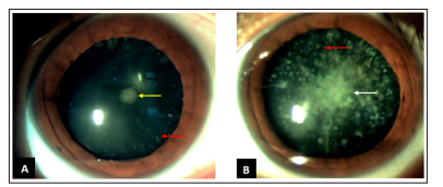
Figure 1: Cerulean Cataract. (A, B) Multiple Opacities spread throughout the Cortex of lens (Red Arrow) these Opacities are radially oriented in the center of the left eye (White Arrow). Note the presence of posterior Subcapsular Cataract in the Right Eye (Yellow Arrow)
Acknowledgements: Published with the consent of the patient.
Conflict of interest: None declared.