Author(s): Sialiti Sanae*, A Khairi, C Hmidi, K Znati, S El Mazouz and K Senouci
Desmoplastic trichoepithelioma is a rare benign tumor emerging from the hair follicle. Here, we report an unusual nasal location of a desmoplastic trichoepithelioma in a female patient.
A 55-year-old female patient with no medical history, presented with solitary facial tumor evolving for 3 years without functional signs, but with major aesthetic discomfort. Medical examination found a flesh-colored soft tumor of 3 x 2cm, located on the left lateral aspect of the nose (Figure 1). There were no other lesions on the face and scalp, nore regional lymphadenopathy. Surgical excision was performed. Histopathological study showed a well-defined tumor proliferation, made up of lobules and clumps surrounded by a retraction slit at the periphery (Figure 2). The tumor cells were basaloid with reduced cytoplasm and regular monomorphic nuclei without atypia or mitosis, as for the stroma, it was fibrous, abundant and cellular, which gave the tumor a double component; epithelial and mesenchymal. CD 34 positive cells surrounded the tumor mass. These features were suggestive of desmoplastic trichoepithelioma. The patient subsequently underwent reconstruction using a glabellar flap with good clinical outcome and no recurrence.
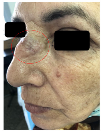
Figure 1: Flesh-colored nodular tumor of the upper third of the nose
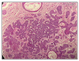
Figure 2: Hematoxylin Eosin Stain: histological aspect of desmoplastic trichoepithelioma showing narrow strands of basaloid cells with fibrous stroma
Desmoplastic trichoepthelioma (DTE) is a rare and benign adnexal tumor of follicular origin fisrt described in 1970. It mainly reported in young adults of 45 year old with a female preponderance [1].
Clinically, this tumor most commonly occurs on sun-exposed areas, particularly the face, rare cases on nose were reported [2]. It is presenting as a painless and well circumscribed nodular lesion ,flesh-couloured and consisting of some lobules, without any change for several years. It’s distinguished by its aesthetic high impact, especially if it is located on the face. It should also be noted that occurrence on the scalp, back or trunk is unusual and poorly reported.
DTE may be difficult to distinguish from other skin tumors, particularly syringoma, skin metastases of breast cancer, scleroderma basal cell carcinoma and microcystic adnexal carcinoma. Histopathological findings correspond to symmetrical tumor covering the entire dermis, not connected to the surface and without deep invasion. It’s consisted of small aggregationsof basaloid cells and numerous small epithelial cysts without peripheral clefting, and stromal cells around cellular islands stain positively with CD34 [3]. Once properly diagnosed, it can be surgically removed with no risk of reccurence.