Author(s): Abdullaiev RY*, Grechanyk OI, Lurin IA, Gumeniuk KV, Posokhov MF and Slesarenko DA
Objective: The present study aims to evaluate the possibilities of duplex ultrasound in the di-agnosis of damage to the large vessels of the neck in combat trauma.
Method: The present study included 59 wounded as a result of mine-explosive, shrapnel ef-fects on the neck. Visualization of large vessels of the neck was carried out using duplex ul-trasound using a linear transducer in the frequency range of 5-10 MHz on Philips HD-11. The p-value <0.05 was regarded as having a significant difference.
Results: Damage to the common carotid artery was recorded in 43 cases, jugular vein - in 39, internal carotid artery - in 24, external carotid artery - in 23, subclavian artery - in 25 cases, subclavian vein - in 25 cases, vertebral artery - in 15 cases, respectively. Types of vascular in-jury included arterial or venous thrombosis, intima detachment with flap, pseudoaneurysm, and arteriovenous fistula. The thrombosis was recorded in 103 cases, detachment of the inti-ma - in 65 cases, pseudoaneurysm - in 26, arteriovenous fistula- in 14 cases, respectively.
Conclusions: Duplex ultrasound allows you to identify damage to all large vessels of the neck received during a combat injury. Thrombosis of the jugular and subclavian veins signifi-cantly predominates over other types of vascular damage (P<0.001). Among the damage to the arteries, the leading place is occupied by detachment of the intima of the common carotid artery (P<0,001).
The neck is a relatively unprotected part of the human body and includes many vital struc-tures, in particular large vessels that supply blood to the brain
According to Verdonck P (2016) in 5% of cases of injury to this body part, only soft tissues are damaged [1]. At the same time, with a neck injury, blood vessels are damaged quite often, and this often leads to mortality and disability [2].
The incidence of vascular injury varies depending on the criteria used and the sensitivity of the diagnostic method used. In CT angiography in patients with a closed head and neck trauma, damage to the carotid artery is detected in 1.07%, vertebral artery in 0.53% of cases, and with fractures of the cervical spine, damage to the vertebral arteries reaches up to 53% [3,4]. Early diagnosis and treatment are critical to reduce neurovascular complications. Digital angiography is considered the gold standard for diagnosing vascular damage in head and neck trauma. Although the effectiveness of the method in the diagnosis of vascular damage is controversial [5,6]. Multidetector computed tomography allows one-stage assessment of dam-age to bone structures and assessment of the state of blood vessels in the angiography mode [7,8].
Types of neck vessel injury include wall dissection with pseudoaneurysm formation, intimal avulsion, arterial or venous thrombosis, arteriovenous fistula formation, and complete rupture. Carotid jugular arteriovenous fistula most often results from blunt trauma to the neck. It is rarely detected in the acute phase after injury, and is very prone to complications such as severe heartfailure, atrial fibrillation, and distal embolization. The presence of a continuous sensation of pressure in the neck, interruptions in the work of the heart, slowing of the heart rate are suspicious clinical symptoms for the presence of an arteriovenous fistula [9].
CT scan may demonstrate expansion of the internal jugular vein due to high flow velocity from the adjacent carotid artery. CT angiography can detect intra-jugular contrast coming from the carotid artery in the arterial phase, allowing the diagnosis of an arteriovenous fistula. One study showed that pseudoaneurysm formed after trauma in 35.7% of cases, after endarterectomy in 35.7% of cases, and due to atherosclerosis in 28.6% of cases [10].
A pseudoaneurysm forms when a disruption in the wall of blood vessels causes blood to pool between the layers of the wall. A true aneurysm is characterized by bulging of all 3 layers of the blood vessel wall. Pseudoaneurysms generally have a high risk of rupture, which is related to the amount of blood flow between the vessel wall and the aneurysm wall [11].
Carotid injury most often results from penetrating neck trauma and carries a high risk of mor-tality. A classification of damage to the carotid artery from degree I (minimal damage to the intima or its unevenness) to degree V (complete rupture of the vessel) has been proposed [12]. In such situations, accurate diagnosis of the nature of the injury is essential. Bedside ultra-sound in color Doppler mode is especially useful in cases where catheter angiography is not possible in unstable patients [13,14]. Doppler ultrasound can record blood flow from the carotid artery to the jugular vein and spectrum arterialization within the jugular vein [15].
In combat operations, the most common causes of damage to large vessels of the neck are mine-explosive, less often bullet impacts. The study of the features of damage to the vessels of the neck using duplex ultrasound, given the non-invasiveness, mobility and information content of the method, is an urgent task.
The aim of the study was to evaluate the role of duplex ultrasound in the diagnosis of damage to the large vessels of the neck in combat trauma.
A retrospective analysis of the results of duplex ultrasound scanning was carried out in 63 wounded soldiers aged 21-37 years who were treated in a military hospital. Various imaging methods were used in complex diagnostics. Duplex ultrasound was performed using a linear transducer in the frequency range of 5-10 MHz on Radmir Pro-30 and Philips HD-11.
Correlation relationships were analyzed by using Pearson correlation coefficient. The p-value <0.05 was regarded as having a significant difference. For categorical variables Fisher exact test was used.
Types of wounds included mine-explosive, gunshot fragmentation, gunshot bullet, blast wave (Figure 1). Damage to the large vessels of the neck due to shrapnel wounds was 41.3%, mine-blast injuries - 31.5%, and other causes - 27.2%, respectively.
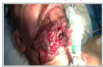
Figure 1: Photo of a fragment wound of the anterior neck with damage to large vessels
Damage to the common carotid artery (CCA) was recorded in 43 cases, jugular vein (JV) - in 39, internal carotid artery (ICA) - in 24, external carotid artery (ECA) - in 23, subclavian ar-tery (RCA) - in 25 cases, subclavian vein (SCV) - in 25 cases, vertebral artery (VA) - in 15 cases, respectively. Types of vascular injury included arterial or venous thrombosis, intima de-tachment with flap, pseudoaneurysm, and arteriovenous fistula (AVF). As can be seen from Table 1, the total number of injuries of the neck vessels in 63 wounded was 194 - of which thrombosis was recorded in 103 cases, detachment of the intima with the appearance of a flap - in 65 cases, pseudoaneurysm - in 26 (Table 1).
As can be seen from Table 1, CCA thrombosis was recorded in 11 (25.6±6.7%) cases, intimal detachment - in 23 (53.5±7.6%) cases, pseudoaneurysm - in 9 (20.9±6.2%) %), ICA - in 7 (29.2±9.3%), 13 (54.2±10.2%), 4 (16.6±7.9%) cases, ECA - in 9 ( 39.1±10.1%), in 12 (52.2±10.4%), in 2 (8.7±5.9%) cases, SCA - in 6 (24.0±8.5%) ), in 8 (32.0±9.3%), in 11 (44.0±9.9%) cases, VA in 6 (40.0±12.6%), in 9 (60.0%) ±12.6%) cases, respectively. Only thrombosis was noted in the jugular and subclavian veins - in 39 (100.0±1.6%) and 25 (100.0±2.0%) cases, respectively. Thrombosis of the jugular and subclavian veins was signifi-cantly more common than intimal detachment and pseudoaneurysm (P<0,001).
| Types of vascular damage |
Damaged vessels | ||||||
|---|---|---|---|---|---|---|---|
| CCA | VJ | BCA | HCA | пкА | пкв | пА | |
| 1 | 4 | 2 | 3 | 5 | 6 | 7 | |
| n=43 | n=39 | n=24 | n=23 | n=25 | n=25 | n=15 | |
| Throm-bosis n=103 |
11 25,6±6,7% |
39 100,0±1,6% |
7 29,2±9,3% |
9 39,1±10,1% |
6 24,0±8,5% |
25 100,0±2,0% |
6 40,0±12,6% |
| Detach-ment of the intima n=65 |
23 53,5±7,6% |
- | 13 54,2±10,2% |
12 52,2±10,4% |
8 32,0±9,3% |
- | 9 60,0±12,6% |
| Pseudoaneurysm n=26 |
9 20,9±6,2% |
- | 4 16,6±7,9% |
2 8,7±5,9% |
11 44,0±9,9% |
- | - |
Note: CCA - common carotid artery; ICA - internal carotid artery; ECA - external carotid artery; JV - jugular vein; SCA - subclavian artery; SCV - subclavian vein; VA - vertebral ar-tery
As can be seen from the table 1, thrombosis occurred in 103 cases, including CCA in 11 (10.7 ± 3.1%) cases, ICA in 7 (6.8 ± 2.5%) cases, NCA in 9 (8, 7±2.8%), JV - in 39 (37.9±4.8%), RCA - in 6 (5.8±2.3%), PCV - in 25 (24.3±4.2%) ) and PA - in 6 (5.8±2.3%) cases, respec-tively. Thrombosis of the jugular and subclavian veins with high (P<0.001) significance was more common than other vessels (Figure 2,3).
Each wounded had damage from 2 to 4 vessels in different combinations - 29 of them had 2 vessels each (54 thromboses + 32 intimal detachments + 12 pseudo aneurysms), 19 had 3 ves-sels each (37 thromboses + 19 intimal detachments +9 pseudo aneurysms + 6 arteriovenous fistulas) and in 12 had 4 vessels each (12 thromboses + 14 intimal detachments + 5 pseudo aneurysms + 8 arteriovenous fistulas).
The results of duplex ultrasound in assessing the frequency of occurrence of intimal detach-ment are presented in table 2. Intimal detachment of the large arteries of the neck was noted in 65 cases - of which CCA in 23 (35.4±5.9%) cases, ICA - in 13 (20, 0±5.0%), ECA - in 12 (18.5±4.8%), SCA - in 8 (12.3±4.1%) and VA - in 9 (13.8±4.3 %) cases, respectively. CCA intimal detachment was recorded significantly more often than ICA (P<0.05), ECA (P<0.05), SCA (P<0.01) and VA (P<0.01).
| Detachment of the intima (n=65) | ||||
|---|---|---|---|---|
| CCA | ICA | ECA | SCA | VA |
| 1 | 2 | 3 | 4 | 5 |
| 23 35,4±5,9% P 1-2<0,05 P 1-3<0,05 P 1-4<0,01 P 1-5<0,01 |
13 20,0±5,0% | 12 18,5±4,8% | 8 12,3±4,1% | 9 13,8±4,3% |
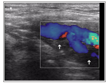
Figure 2: Thrombosis of the Internal Carotid Artery as a Result of a Mine-Explosive Injury. In Color Doppler Mode, there is no Blood Flow in the Lumen of the Internal Carotid Artery (Arrows)
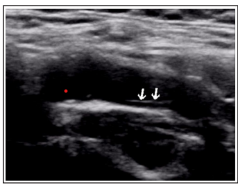
Figure 3: Detachment of the Intima of the Internal Carotid Artery (Arrows)
Pseudoaneurysm was diagnosed in 26 cases, of which 9 (34.6±9.3%) cases of OSA, 4 (15.4±7.0%) cases of ICA, and 2 (7.7±5.2%) cases - ECA and in 8 (30.8±9.1%) - RCA, re-spectively. Pseudoaneurysm of the CCA and RCA with minimal significance (P<0.05) was noted more often than the ECA (Figure 4).
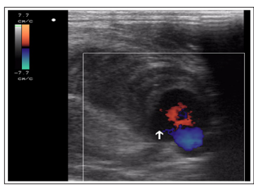
Figure 4: Formation of Common Carotid Artery Pseudoaneurysm as a Result of a Mine-explosive shrapnel wound. Arrow indicates false canal thrombosis
Arteriovenous fistula was formed in 14 cases - in 8 of them (57.1±13.2%) between the com-mon carotid artery and jugular vein, in 6 (42.9±13.2%) cases between the subclavian artery and subclavian vein (Figure 5)
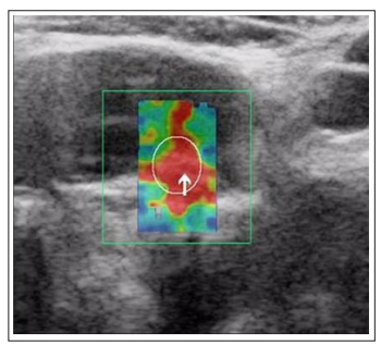
Figure 5: Formation of an arteriovenous fistula between the common carotid artery and the internal jugular vein (arrow)
We have analyzed the frequency of occurrence of various types of damage to the common carotid artery (Table 3). As can be seen from the table, intimal detachment was significantly more common than thrombosis (P<0.05), pseudoaneurysm (P<0.001) and arteriovenous fistula (P<0.001).
| Common carotid artery (n=51) | |||
|---|---|---|---|
| Thrombosis | Detachment of the intima |
Pseudoaneurysm | Arteriovenous fis-tula |
| 1 | 2 | 3 | 4 |
| 11 21,6±5,8% |
23 45,1±7,0% P 2-1<0,05 P 2-3<0,001 P 2-4<0,001 |
9 17,6±5,3% | 15,7±5,1% P 4-3<0,05 |
It is known that in the neck area there are many vital organs - large blood vessels supplying the brain, spinal cord, trachea, esophagus. Any foreign bodies that are directed to this area of the body during combat operations as a result of mine-explosive, fragmentation, bullet impact can easily injure the tissues of the neck, since it is less protected [1]. Types of damage to large vessels can vary from local detachment of the intima to complete rupture [2]. The latter in most cases leads to immediate death.
Previously published works present data on damage to soft tissues and large vessels of the lower extremities in multiple injuries of various origins, in particular, combat injuries [15,16]. Published works on the diagnosis of post-traumatic injuries of soft tissues and bone structures mainly present the results of computed tomography [17].
We have found that thrombosis of the jugular and subclavian veins are significantly more common than those of arterial vessels. Among the latter, the common carotid artery is more often affected, and intima detachment occupies a leading place among the various types of damage.
Duplex ultrasound allows you to identify damage to all large vessels of the neck received dur-ing a combat injury. Thrombosis significantly predominates over other types of vascular dam-age. They often occur in the jugular and subclavian veins. Among the damage to the arteries, the leading place is occupied by detachment of the intima of the common carotid artery. Arteriovenous fistula is much less common.
The authors declared no potential conflict of interest with respect to the research, authorship, and/or publication of this article.
