Author(s): Kaamraan Islam and Omar Islam*
Via a pictorial review, we will discuss the complex anatomy and important function of the kidney as it relates to urine formation and excretion. We will introduce the anatomy and function of the renal lobe and review the histology of the renal cortex and medulla, renal tubule, renal papilla, and renal pelvis. We will discuss renal filtration capacity and review the pathway of renal excretion through the ureters, urinary bladder, and urethra.
First, let’s discuss an analogy. Imagine we are boiling some water to make pasta. 10 minutes later, hopefully we’ll have cooked pasta which is our desirable substance, and some dirty water, which is our undesirable substance. We have one problem, which is that the cooked pasta and dirty water are mixed together in the same pot! What is the solution? One solution is to use a strainer. A strainer would allow us to keep the desirable substance which is the cooked pasta, while removing the dirty water or the undesirable fluid. The undesirable fluid would then be directed down the drain [1-3].
Now you may be wondering, why we are talking about pasta. Weren’t we supposed to be learning about the urinary system? Well actually, we were learning about the urinary system. It turns out that straining pasta and releasing the dirty water down the drain is very similar in nature to the urinary system (Figure 1).
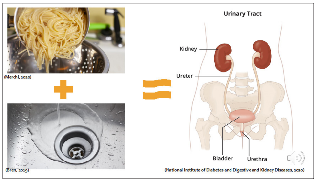
Figure 1: Analogy
The purpose of the urinary system is to filter blood. Toxins from the blood are made into a substance called urine. You can see a representation of the kidneys and ureters (Figure 2). These are some of the key players in system. This task can be thought of similar to straining pasta.
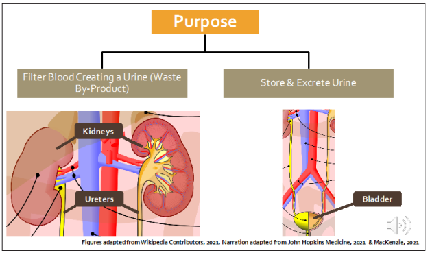
Figure 2: Purpose of the Urinary System
The urinary system is also responsible for storing and releasing urine [4,5]. The bladder as depicted in the diagram is also an important part of the system. This task can be thought of similar to pouring dirty water down the drain.
Now that we have an overview of the urinary system, let’s learn about how blood is filtered and how urine is formed. But first, we need to spatially orient ourselves with the kidneys and identify some important structures [6-15].
The kidneys are located on either side of the spin in the abdominal cavity between the thoracic and lumber vertebrae. Here is a coronal cross section of the right kidney (Figure 3). It is important to be able to identify two main parts in this diagram. The first structure we should know is the hilum, which is the surface where renal arteries and veins enter and exit the kidneys. The blood then travels to the renal lobes which appear dark red, and shell shaped in this diagram. The renal lobes are the most important part of the kidney, as blood is filtered to make urine in this region.
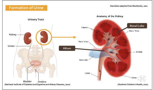
Figure 3: Anatomy of the Kidneys
The renal lobe consists of two main parts (Figure 4): the renal cortex which makes up the outer layer of the kidney and lobe, and the renal medulla which makes up the inner region deep to the cortex [16-18]. Together, the renal medulla and the renal cortex filter blood to make urine.
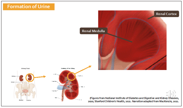
Figure 4: Anatomy of the Renal Lobe
The functional unit of the kidney is called the nephron. The nephron (Figure 5) filters blood and produces urine. At first it may look complicated, but it is relatively straightforward. The content above the horizontal brown line in the diagram is situated in the renal cortex, while the region below the brown line is in the renal medulla.
Next, we will focus on the structure at the top left of the diagram known as the renal corpuscle.
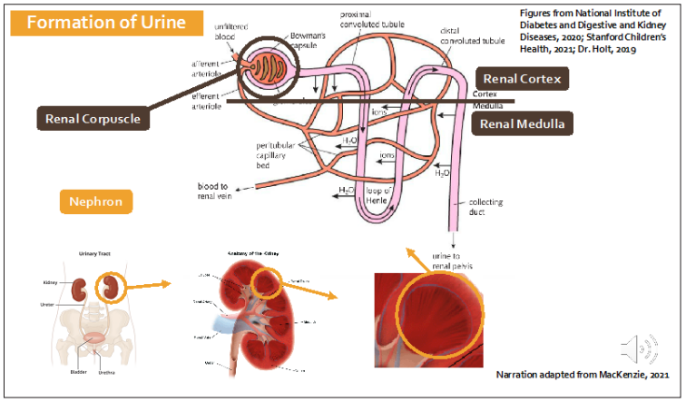
Figure 5: Histology and Function of the Renal Cortex and Medulla
The renal corpuscle is where the first step in the formation of urine takes place. Blood enters the kidney and flows into the glomerular capillaries forming a structure called the glomerulus (Figure 6) which is essentially a bundle of capillaries. The glomerular capillaries have fenestrations, or small holes that allow ions, water, and other waste products to diffuse out of the blood into Bowman’s space. This is the white region between the red capillaries and the pink structure. The filtrate then enters the Bowman’s Capsule which is the pink structure in this diagram.
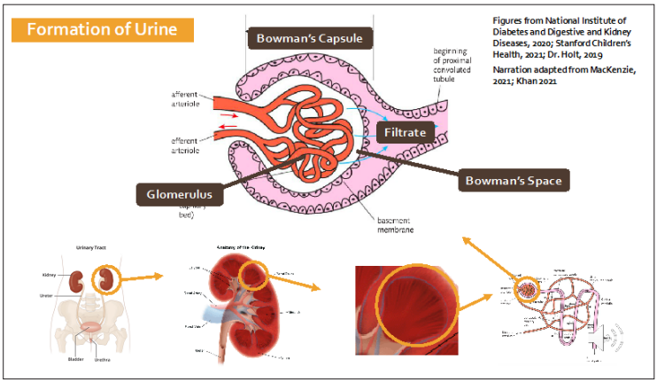
Figure 6: Histology and Function of the Renal Corpuscle
Up until now, we have described the first step in the formation of urine. The second step involves the renal tubule. The main purpose of the renal tubule is to conserve water from the newly formed filtrate. There are four main regions that we need to understand (Figure 7).
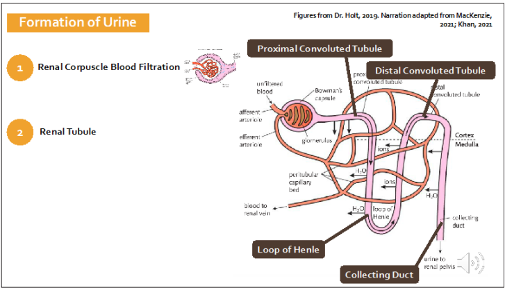
Figure 7: Histology and Function of the Renal Tubule
The first region is called the proximal convoluted tubule. It is an extension of the Bowman’s capsule. Valuable items from the filtrate that had been filtered out of the blood such as glucose and sodium are actively reabsorbed back into the blood stream in this section.
The next, and arguably most important part of the nephron is called the loop of Henle. It actively pumps out salt from the filtrate. This causes water to leave the loop of Henle. So essentially, the loop of Henle conserves water.
The distal convoluted tubule performs similar functions to the proximal convoluted tubule as it reabsorbs valuable molecules. By the time the valuable molecules have been reabsorbed, the filtrate enters the collecting ducts which can reabsorb more water if necessary, with the help of anti-diuretic hormone.
The newly formed filtrate then travels from the collecting duct into the renal papilla (Figure 8). At this point, the filtrate can be called urine.
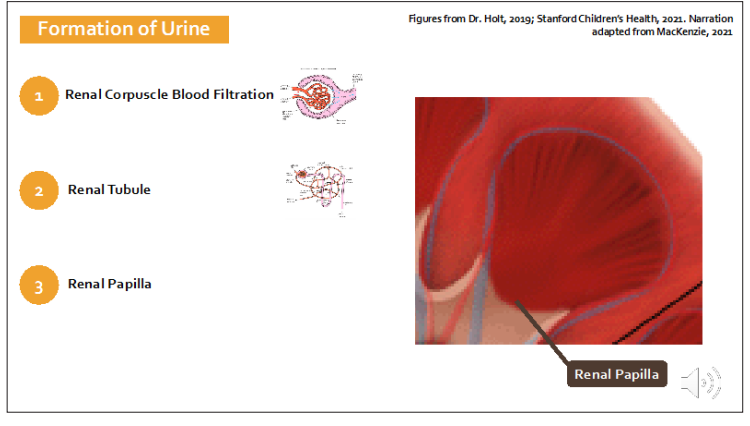
Figure 8: Histology and Function of the Renal Papilla
The urine then flows from renal papilla into the minor calyces, which become major calyces and then flow into the renal pelvis (Figure 9). The renal pelvis serves as a catchment area for the newly formed urine from all the lobes in the kidney. The renal pelvis then drains urine into the ureter.
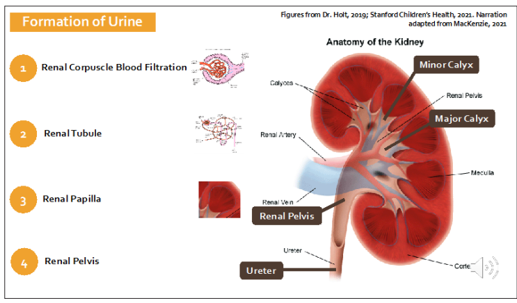
Figure 9: Histology and Function of the Renal Pelvis
The kidneys can filter up to 1.1 litres of blood per minute. That’s the same amount of fluid as in a bottle of coke (Figure 10).
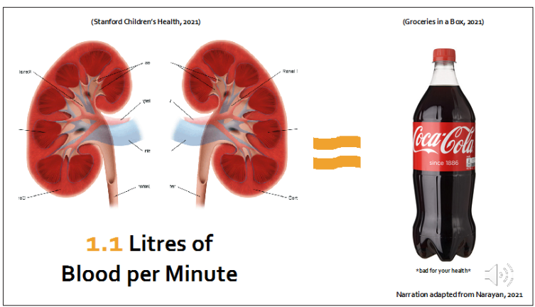
Figure 10: Filtration Capacity
Now that we understand how urine is formed, let’s briefly discuss how it is stored and released from the body. This process is a lot less complicated than the formation of urine.
The first step in this process involves the ureters (Figure 11). As depicted in the diagram, the ureters are muscular tubes that connect the kidneys to the urinary bladder. They connect on the medial side of both kidneys and to the posterior of the urinary bladder. The purpose of the ureters is to transport newly formed urine from the kidneys to the urinary bladder.
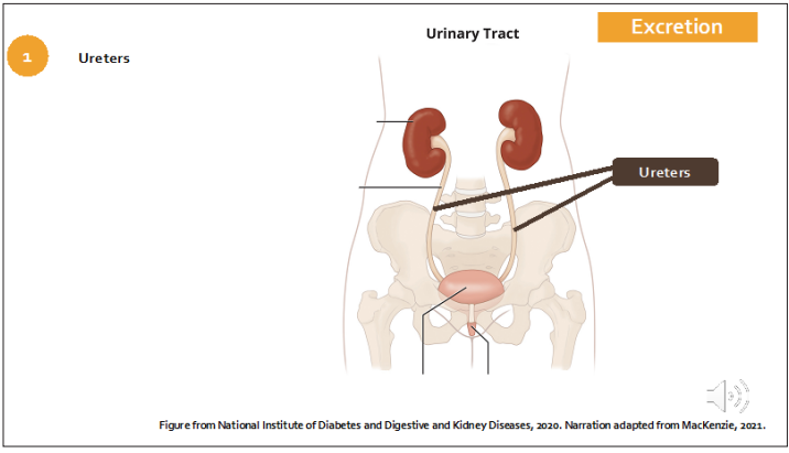
Figure 11: Excretion Ureters
The second step involves the urinary bladder (Figure 12). The urinary bladder serves as a temporary storage unit for urine.
Urine enters the urinary bladder through the ureters. You can see the left ureteric opening or where the left ureter releases urine into the bladder on the diagram.
The trigone, which as you may be able to guess, forms a triangle with the right and left ureteric openings superiorly, and the internal urethral opening inferiorly. The trigone funnels urine into the internal urethral opening.
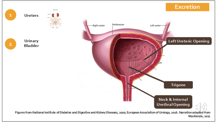
Figure 12: Excretion Urinary Bladder
The average capacity of the adult human urinary bladder (Figure 13) is up to 2 cups of water
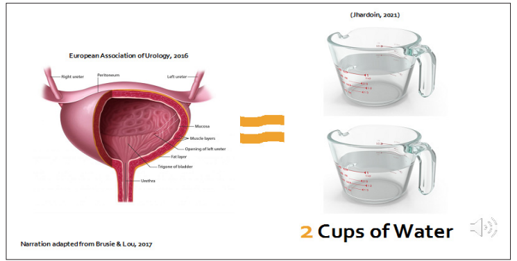
Figure 13: Excretion Urinary Bladder capacity
The third step in the excretion of urine involves the urethra (Figure 14). The internal urethral opening directs urine into the urethra, which is the third site in the urinary excretion process [19, 20]. The purpose of the urethra is to release urine from the bladder through an opening called the external urethral meatus. This diagram depicts the female urethra, which is approximately 3-5 cm long.
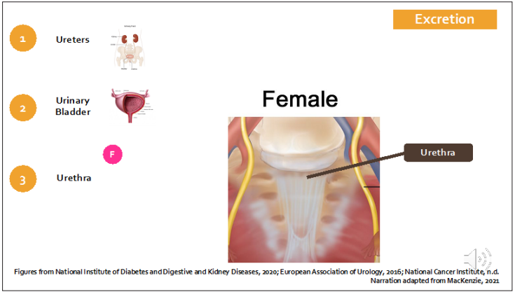
Figure 14: Excretion Female Urethra
The male urethra (Figure 15) is significantly longer than the female urethra measuring approximately 20 cm. It is divided into three regions: the prostatic urethra, the membranous urethra, and the penile or spongy urethra.
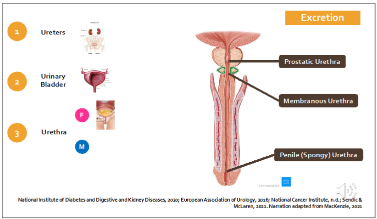
Figure 15: Excretion Male Urethra
The fourth and final step of urinary excretion is micturition, or the act of urination. In this step, the external urinary sphincter relaxes which allows urine to travel from the bladder into the urethra to be excreted (Figure 16).
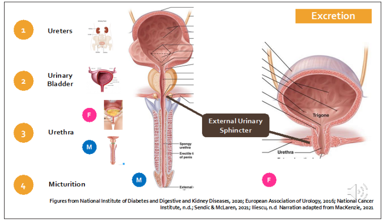
Figure 16: Excretion Micturition
There are similarities between straining pasta and the urinary system (Figure 17). The pasta strainer acts like a kidney. It separates the desirable from the undesirable substances. The sink is like the bladder. It holds the undesirable substance. And the drain is like urethra. It releases the undesirable substance [21,22]. The analogy of straining pasta illustrates the fundamentals of the complex anatomy and important function of the kidney as it relates to urine formation and excretion.

Figure 17: Summary
The authors declare no conflict of interest.
The data supporting this SYSTEMATIC REVIEW are from previously reported studies and datasets, which have been cited.
The authors declare no financial support for the research,authorship, or publication of this article.