Author(s): Mansour Nacouzi
Granular cell tumor or Abrikossoff’s tumor is an infrequent tumor that can arise in most organs, and especially in the ENT area. It is a usually benign neoplasm, that can lead to a misdiagnose of malignancy. It affects both sex, between the fourth and the sixth decade. We present in this report a case of a 14 years old girl with a slowly growing lesion on the right lateral border of the mobile tongue. The biopsy showed a proliferation of large cells with a granular cytoplasm that expressed two immunohistochemistry markers: CD68 and S100 antibodies. Surgical resection was completed with a one centimeter margin. The rare issue about this case is the age of presentation: the age of the patient is 14, whereas this tumor usually affects adult patients.
Abrikossoff’s tumor or granular cell tumor is an infrequent, usually benign neoplasm, first described by the Russian pathologist Abrikossoff in 1926. Most cases affect deep dermis and subcutaneous tissue, particularly of the head and neck, trunk, and proximal extremities. The most common site is the tongue, representing 25% of cases, following by the breast (5-15%). Visceral involvement of the gastrointestinal tract and respiratory tract is also common. These tumors usually occur in the fourth to sixth decade of life, and they are generally reported to be about 2-3 times more common in females than males [1]. The estimated incidence of oral GCT is approximately 1/1.000 000 per year. Malignant granular cell tumor is very rare and occurs with a female predominance.
Granular cell tumors are a unique, nodular, firm masses, often with an overlying epithelial hyperplasia. that can lead to a misdiagnose of a malignant epithelial tumor. On sectioning, the tumor has a yellowish finely granular texture. In this article, we will discuss an unusual case of a 14 years old girl with a tongue GCT.
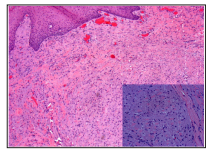
We present a case of a 14 years old girl, who presented in Pasteur Hospital in Colmar-France, for a painless tongue lesion, slowly growing for over a year. (Figure: 1) Physical examination showed an indurated and sensitive nodular lesion of the middle third of the right lateral border of the tongue. Neck examination revealed no adenopathy. A biopsy was performed. A proliferation of large eosinophilic cells with a finely pale pink and foamy granular cytoplasm, was identified in the chorion. The mucosa above the lesion showed an acanthosis, without dysplasia. (Figure: 2). No pathogenic element were identified with a PAS staining.
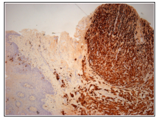
An immunohistochemistry technique showed the tumor cells stain with CD68 and S100 .(Figure: 3)
These findings on H&E stain and immunohistochemistry confirm the diagnosis of Granular cell tumor.
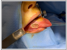
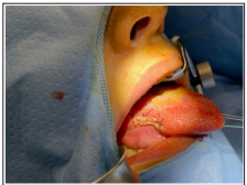
A total excision under general anesthesia, was realizes with a one centimeter margin. (Figure: 4,5)

Figure 1: Painless firm tongue lesion, slowly growing for over a year

Figure 2: The squamous epithelium of the tongue shows hyperplasia, overlying a proliferation of large eosinophilic cells, with granular cytoplasm

Figure 3: On immunohistochemistry, S100 marker is diffuse and strongly positive in Granular cell tumor

Figure 4: Directly after the resection of the tongue tumor

Figure 5: After suturing the mucosa of the tongue
Microscopically, granular cell tumor has ill-defined borders and is composed of sheets, nests, and trabeculae of large, monotonous epithelioid to polygonal cells with intensely eosinophilic, granular cytoplasm. Nuclei are usually centrally situated and range from uniformly small and mildly hyperchromatic to large and vesicular with distinct nucleoli. The granular appearance of the cytoplasm due to massive accumulation of lysosomes including large intracytoplasmic granules highlighted by clear haloes, that are filled with PAS+, diastase resistant granules. (Malignant granular cell tumors are rare and their features vary from overtly sarcomatous to morphologically bland on rare occasions).
Rare examples of malignant granular cell tumour have also been described. Histological features associated with malignancy include increased cellularity, prominent spindling, high nuclear/ cytoplasm ratio, vesicular nuclei with prominent nucleoli, marked pleomorphism, increased mitotic activity, and geographical necrosis.
Granular cell tumors are generally reactive for S100, SOX10, nestin, inhibin, and calretinin. Tumors are also positive for CD68, CD63, and NSE. MITF and TFE3 show diffuse nuclear reactivity in most cases, but HMB45 is uniformly negative and only very rarely is there focal reactivity for melan-A. lmmunohistochemical studies suggest differentiation consistent with an origin from Schwann cells.
The histological desirable diagnostic criteria are nests and sheets of epithelioid to polygonal cells, abundant intensely eosinophilic, granular cytoplasm, positive for S100 on immunohistochemistry. On the surgical level: many articles in the literature interest Abrikossoff tumor in the ENT area. To begin with a case series of 5 patients in Greece, one of them is a 59 years old men with a tongue lesion that turned out to be a granular cell treated with an en-bloc surgical resection with no recurrence after 4 years of follow up [2].
Another case diagnosed in Napoli, Italy of a 36 years old female presenting a granular cell tumor of 17 millimeter diameter. Immunohistochemistry confirmed the diagnosis with S100-positive cells containing Cd68-reactive granules. A surgical excision was done with a minimum margin of 0.3 mm from the deep structures. A one year follow up showed absence of recurrence [3].
Another case in Portugal is about a 43-year-old female patient, referred to the stomatology consultation for the observation painless and slow-growing lesion of the tongue. It was about 1.5 x 0.9 x 0.4 cm in diameter, located at the posterior limit of the middle third of the dorsum of the tongue. It was surgically removed with margins. The follow up did not show any signs of recurrence one year after the surgery.
The majority of Granular cell tumor patients found in the literature belong to the adult population. However, our case is an adolescent patient with 14 years of age. That makes this case report interesting [4].
Granular cell tumor usually occurs more in men than women, in their fourth to sixth decade. This is a case of a 14 years old girl, who was diagnosed with granular cell tumor of the lateral border of the tongue.
The pathology diagnosis was made with H&E staining showing large cells with granular cytoplasm, and CD68+ and S100+ immunostaining profile. The surgical resection was made with one centimeter margins that turned to be 1mm negative margins on pathologic report.
