Author(s): El Baroudi Taieb*, Belghmaidi Sarah, Hajji Ibtissam and Moutaouakil Abdeljalil
Retinoblastoma is the most common intraocular cancer of childhood. We report the case of a 3-year-old child with unilateral leucocoria noticed by parents
who revealed a retinoblastoma. Leucocoria can also indicate other vision threatening conditions : Coats’ disease, cataract, toxocariasis, retinopathy of
prematurity for which prompt medical attention is needed.
Retinoblastoma is the most common intraocular cancer of childhood. It is initiated by mutation of the RB1 gene, which was the fi rst described tumour-suppressor gene [1-3]. Incidence of retinoblastoma is constant worldwide at one case per 15 000- 20 000 livebirths, which corresponds to about 9000 new cases every year [4].
We report the case of a 3-year-old child with unilateral leucocoria noticed by parents figure 1 showing an unilateral left leucocoria. The child benefited from an examination under sedation which showed the presence of a whitish, vascularized and polylobed intra retinal mass with micro-calsifications figure 2. The child also received a mode B ocular ultrasound which showed the presence of an intra vitreous tissue formation with microcalcification figure 3. The MRI confirmed the diagnosis and allowed to show the absence of extension to the sclere or the optic nerve. The examination of the contralateral eye was without particularity. The extension assessment was without particularity. In front of these clinical and radiological elements the child benefited from an urgent ennucleation. The pathology study confirmed the diagnosis of retinoblastoma and the absence of tumor invasion of the scleral and optic nerve.
Patient consent confirmation: I declare on my honor that clear and informed consent has been obtained from the child’s parents to publish the findings of this case study.
Conflicts interest: We have no conflicts of interest to disclose.
Leucocoria is the most common initial sign of retinoblastoma and is first apparent when the tumour is still contained within the eye. The attending physician and the parents must be attentive to any change in the child’s pupil glow.
Leucocoria can also indicate other vision threatening conditions : Coats’ disease, cataract, toxocariasis, retinopathy of prematurity for which prompt medical attention is needed.
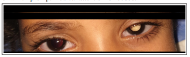
Figure 1: Showing an unilateral left leucocoria
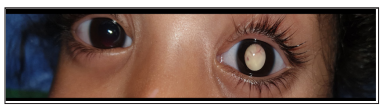
Figure 2: Showing a whitish, retrocrystalline, polylobed and vascularized mass
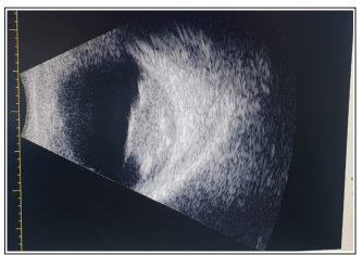
Figure 3: Mode B ocular ultrasound which shows the presence of an intra vitrial tissue formation with micro-calcifications