Author(s): Anubhav Chauhan*, Deepak Kumar Sharma and Anchit Wapa
A 20-year-old male visited our hospital for routine ocular examination. He was a known case of Neurofibromatosis Type 1(NF1). There was no other significant history. His visual aquity was 6/6 in both the eyes. Bilateral fundus examination, colour vision, intraocular pressure, and ocular movements were within normal limits. Slit lamp examination revealed iris hamartomas (lisch nodules) in both the iris (figure 1 and 2). He was advised a regular ophthalmology followup.
Karl Lisch, an Austrian ophthalmologist was the first person who described the association of NF1 with these iris hamartomas. These nodules are 1-2 mm in size, yellow brown coloured, and dome shaped and present over the iris surface [1]. NF1 is a neuro-cutaneous disorder with lisch nodules, optic nerve glioma, sphenoid dysplasia etc. being important ocular findings of this disease [2]. Lisch nodules are ocular pathognomonic markers of NF1 [3]. They are asymptomatic and do not require treatment [4].
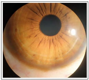
Figure 1: Lisch Nodules-Right Eye
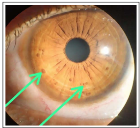
Figure 2: Lisch Nodules-Left Eye
Conflicts of Interest: The authors declare that they have no competing interest.
Financial Disclosure: The authors have no proprietary or commercial interest in any material discussed in this article.