Author(s): Dessalew Habte*, Dessalew Tamir and Tadesse Tilahun
Erysipelas is an infectious disease seen mainly in growing pigs and characterized clinically by fever, arthritis, skin lesions and sudden death. The disease may be acute, subacute, or chronic. Swine erysipelas is a disease caused by a specific micro organ Erysipelothrix rhusiopathiae whic h is a zoonotic ubiquitous gram-positive bacterium that causes erysipelas in swine, mammals, birds and erysipeloid in humans. People in contact with animals, animal products or animal wastes are at greatest risk. The acute form of swine erysipelas may have been confused for other diseases in pigs which are characterized by acute symptoms such as sudden death (for example, African swine fever). It can be diagnosed by its clinical signs, necropsy findings, bacteriology, antimicrobial response, molecular and serological examinations. It is recommended to increase awareness of the disease among animal and human practitioners as treatment is easy and available and vaccination is possible. However, the disease is still unknown to local veterinarians, clinical doctors, meat inspectors, butchers and laboratory personnel. Proper hygiene, regular pork inspection, use of protective wear among people working/ in contact with animals should be promoted. The disease causes high economic loss in pig rearing areas and influences the public health being a severe zoonotic disease. So the objective of this review is to create a better understanding of the disease for proper control and prevention of the disease.
Erysipelas, one of the oldest recognized diseases is an infectious disease caused by Erysipelothri x rhusiopathiaeseen mainly in growing pigs and adult swine and characterized clinically by sudden death, fever, arthritis, and skin lesions. The disease may be acute, subacute, or chronic. Up to 50% of pigs in intensive swine production areas are considered to be colonized with E. rhusiopathiae. The causative agent is a bacterium, Erysipelothrixr husiopathiae. The organism commonly resides in the tonsillar tissue. These typical healthy carriers can shed the organism in their feces or oronasal secretions and are an important source of infection for other pigs [2]. The mode of transmission of the disease is by ingestion, natural infection through skin wound, biting flies, intrauterine infection, and soil contaminated with organisms and contaminated water also aids the spread of infection [3].
Disease outbreaks may be acute or chronic, and clinically inapparent infections also occur. Acute outbreaks are characterized by sudden and unexpected deaths, febrile episodes, painful joints, and skin lesions that vary from generalized cyanosis to the often-described diamond skin (rhomboid urticaria) lesions. Chronic erysipelas tends to follow acute outbreaks and is characterized by enlarged joints and lameness. A second form of chronic erysipelas is vegetative valvular endocarditis [4]. The disease can be prevented and controlled by proper management, sanitation and vaccination measures and can also be effectively treated by penicillin but what makes it difficult to control is its occurrence in all ages of pigs in different f arms and the presence of carriers. The disease causes high economic loss in pig rearing areas and influences the public health being a severe zoonotic disease. So the objective of this review is to create a better understanding of the disease for proper control and prevention of the disease [5].E. rhusiopathiae serotypes 1 and 2 are frequently isolated from clinically affected pigs, although other E. rhusiopathiae serotypes have been sporadically associated with clinical disease. While there is no experimental evidence that Erysipelothrix species other than E. rhusiopathiae cause disease in pigs, certain Erysipelothrix species strains have been isolated from clinical cases and from condemned carcasses in abattoirs [9].
Growth of E. rhusiopathiae on nonenriched media produces pinpoint, nonhemolytic colonies after incubation for 24 hr. After 48 hr of incubation, a zone of incomplete hemolysis becomes evident around colonies. The genus of Erysipelothrix is subdivided into two major species: E rhusiopathiae and E. tonsillarum. In addition, there are other strains that constitute one or more additional species known as E species 1, E species 2, E species 3, and E. inopinata. At least 28 different serotypes of Erysipelothrix spp are recognized, and pigs are considered to be susceptible to at least 15. Field cases of swine erysipelas are predominately caused by E. rhusiopathiae serotypes 1a, 1b, or 2 [10].Older colonies can lose cell wall integrity, making them appear to be gram negative or gram variable. The organism grows readily on standard media and in conventional blood culture systems. Both rough and smooth colonies are noted on blood agar. Smears from rough colonies can show granular, nonbranching filaments. Some strains can cause α-hemolysis in 48 to 72 hours. E. rhusiopathiae is distinguished readily from other morphologically similar, gram-positivebacteria (Listeria species and diphtheroids) by an absence of motility, a negative catalase reaction, and hydrogen sulfide (H2S) production. E. rhusiopathiae is oxidase and urease negativ e; it produces acid from glucose and lactose. A growth pattern in gelatin stab cultures that resembles a test-tube brush or a pipe cleaner is highly characteristic [11].
E. rhusiopathiae is regarded as a commensal organism of swine flora and is found in the upper respiratory tract and the intestinal tract of healthy pigs. Greater than 50% of pigs are considered to be carriers. E. rhusiopathiae survives in the soil for prolonged periods and may also be found in other mammalian species including sheep, fish, and birds. E. rhusiopathiae is considered to be a zoonotic agent [12, 13].The infection, usually with serotypes 1a or 2, has also been demonstrated in wild boars, so these should not be forgotten as a reservoir. Morbidity and case fatality rates in pigs vary considerably from area to area. Because of variations in virulence of the particular strain of the organism involved in infection. Sources of infection are from domestic pigs, carrier animals? excretafaeces, urine, contaminated water and feed, body secretions-saliva, nasal [15]. Predisposing factors are age, genetics, immunity, non- infectious disease, tress due to environment or management (nutrition, ambient temperature, and fatigue), worm infestation, concurrent infection and alkaline soil. The incidence of human infection is higher in the summer months. A 4:1 male preponderance may reflect male occupational exposure [16].
Swine less than 3 months or more than 3 years of age are generally least predisposed to SE. The relationship of age to susceptibility may be explained by naturally acquired passive immunity in the young and active immunity following subclinical infection in older animals. Suckling pigs of immune sows are immune to infection for several weeks after birth. The degree and duration of passive immunity are related to the immune status of the sow. Parasitic infestations have been reported to increase the severity of clinical SE. Susceptibility of swine to acute SE can be enhanced by subclinical toxicity from aflatoxin in the feed. Sudden outbreaks of acute SE may be the result of a combination of the susceptibility of the animals and virulence of the causative organism [17].E. rhusiopathiae, a ubiquitous organism, is primarily a pathogen of animals. It has been isolated from wild mammals; various fowl, fish, and shellfish; and from domestic animals, such as pigs, sheep, cattle, and horses. Contaminated soil is thought to be a source. Human infections often are to the result of occupational exposure to infected animals or contaminated animal products. People at risk include slaughterhouse workers, butchers, poultry workers, fishermen, fish marketers, veterinarians, farmers, and housewives [18].
On farms where the organism is endemic, pigs are exposed naturally to E. rhusiopathiae when they are young. Maternalderived antibodies provide passive immunity and suppress clinical disease. Older pigs tend to develop protective active immunity as a result of exposure to the organism, which does not necessarily lead to clinical disease. E rhusiopathiae is excreted by infected pigs in feces and oronasal secretions, effectively contaminating the environment. When ingested, the organism can survive passage through the hostile environment of the stomach and intestines and may remain viable in the feces for several months. Recovered pigs and chronically infected pigs may become carriers of E rhusiopathiae. Healthy swine also may be asymptomatic carriers. Infection is by ingestion of contaminated feed, water, or feces and through skin abrasions [19].Infections of man and animals have been documented from Africa, Australia, several countries in the Americas, Japan, China and throughout Europe. Human disease can originate from an animal or environmental source [20]. Interestingly, outbreaks of vegetative endocarditis can occur in the absence of other clinical signs. In older, susceptible swine, various stresses (heat, aflatoxin, poor nutrition) have been suspected of playing a role in precipitating outbreaks [21].
Recovered pigs and those chronically infected may be carriers of the organism, possibly for life. The mode of entry is by ingestion and through skin abrasions. Following ingestion, the organism most likely enters the body via the tonsils or lymphoid tissue of the GI tract. In small populations kept in back yards, orchards or paddocks there is plenty of opportunity for access by birds and rodents and the persistence of the organism in soil favors persistence of disease [23].
Erysipelas is particularly evident in systems that allow or promote: Contact with bird faeces, mouse contamination, and access to solid muck. In practice, this means that the disease is most prevalent in straw based systems, particularly in open barns (i.e. the supposed welfare friendly pig keeping systems) and tends to peak in the summer months, although can occur at any time [24].Because of human susceptibility, swine erysipelas has some public health significance. Veterinarians in particular are exposed to infection when vaccinating with virulent cultures. It commonly contaminates pig products and therefore is quite a common infection in abattoir workers or butchers or those employed in similar trades. It usually produces a swollen finger and is known as erysipeloid. In this context, there have been recent advances in slide agglutination and latex agglutination tests for rapid diagnosis, which have a good correlation with each other and subsequent culture. Now a PCR identifying four species has been described, principally for use in the abattoir.
Recently a case of endocarditis and presumptive osteomyelitis has been described, so care is needed. Type 21 is recorded as having produced a septicemia in humans [27].Acute infection has septicemia with disseminated intravascular coagulation and hyaline thrombi throughout the body. By four days post infection, the bacteria invade the endothelium, and there is diapedesis of erythrocytes. The purple skin is usually due to congestion and sometimes thrombosis of dermal vessels. Fibrinoid necrosis of vessels may be due to a hypersensitivity (Arthus) reaction. Arteriolar fibrinoid necrosis is thought to be the cause of the ?diamond skin? lesions and may not be present in our piglets due to the rapid nature of the infection which may not have allowed enough time for full hypersensitivity vasculitis to develop. The discoloration of the skin can be used as a prognosticator. Pigs with pink to red skin lesions usually recover, while those with dark red-purple lesions usually die. In acute erysipelas in piglets, the dermal and hypodermal hemorrhage also occurred mostly on the ears and limbs. Muscle degeneration is seen with acute erysipelas, but the locally severe pattern of hemorrhagic infarction seen in one of our pigs is unusual. Less specific lesions can be seen in any organ, with leukothrombi or bacterial emboli. Synovitis may occur in acute or chronic disease. The subacute form is similar but less severe than the acute disease [29].
In chronic arthritic cases, there is a vasculitis with exudation of fibrin into perivascular tissues and joint(s). There also is marked villous proliferation of the synovial membrane. Joint lesions are more proliferative than exudative. Connective tissue formation around joints is stimulated, apparently by perivascular fibrin, and the joint capsule may be thickened. Persistence of inflammation around an affected joint may be from a few living bacteria in the joint or it may be the result of hypersensitivity to remaining bacterial antigen [29,30].Vegetative, valvular endocarditis, another manifestation of chronic erysipelas, is less common than joint involvement. Emboli of E. rhusiopathiaeare believed to cause inflammation of blood vessels within heart valves. In the inflammatory process, the endocardium is breached and bacterial colonies are established at the site. Fibrin is deposited there and slowly organized to form nodular vegetations. Emboli that arise from vegetations can cause sudden death [31].
Acute cases occur after an incubation period of 1-7 days with sudden death with high rise in temperature (104-106°F), stilly gait and get up with difficult suspended bowl material movement. Pigs remain depressed and burrow in the bedding. Conjunctivitis and vomition, anorexia and thirst are common. Slightly pink to dark purple area which raised and firm to touch. Diamond skin lesions from sick pigs often have reddened or cyanotic skin, especially about the ears, snout, jowls, throat and ventral abdomen [33].
Chronic form is the most common form of erysipelas that may follow acute or subacute disease as well as subclinical infection and is characterized most commonly by arthritis. Arthritis mainly involved joints are hock, stifles, knee, and elbow and Losses occur due to lameness, ill-thrift and carcass condemnation. Joints are stiff, enlarged, hot and painful with sloughing of the tip of the tail or tips of the ears can occur but can have other causes. Signs of heart problems due to infection of the heart valves may be evident occasionally and will be most obvious after exertion, which may lead to sudden death. Paraplegia may occur when intervertebral joints are involved or when there is gross distortion of limb joints [34].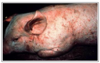
Figure 1: Acute swine erysipelas, skin form [35].
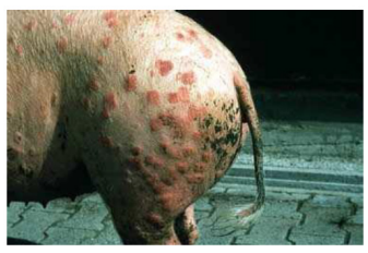
Figure 2: Typical diamond-shaped skin lesions [35].
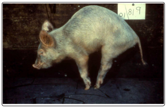
Figure 3: Crippling, irreversible arthritis in a growing pig [35].
In chronic erysipelas, valvular endocarditis is seen as proliferative, granular growths on the heart valves, and embolisms and infarctions may develop. Arthritis may involve joints of one or more legs, and the intervertebral articulations may be involved. Affected joints may be enlarged, with proliferative, villous synovitis and increased viscosity of synovial fluid, inflammatory exudates, and thickening of the joint capsule. Proliferation and erosion of articular cartilage may result in fibrosis and ankylosis of the joint [37].
Isolation of E. rhusiopathiae from blood of affected pigs, especially after enrichment, is possible in acute cases and helps establish a diagnosis. In addition, molecular methods capable of detecting E. rhusiopathiae DNA in affected tissues or blood (i.e., PCR assays) can also be used. Recently, immunohistochemical methods to demonstrate the organisms in formalin-fixed paraffinembedded tissues have become available and are useful in cases when pigs have been treated with antimicrobials before sample submission. A rapid, positive response to penicillin therapy in affected pigs supports a diagnosis of acute erysipelas because of the sensitivity of the organism to penicillin [39].
Chronic erysipelas can be difficult to definitively diagnose. Arthritis and lameness, coupled with the presence of vegetative valvular endocarditis postmortem, may support a presumptive diagnosis of chronic erysipelas. However, these lesions can be caused by other infectious agents. A positive culture of valvular vegetations or demonstration of E. rhusiopathiae DNA in the lesions by PCR is definitive for diagnosing chronic erysipelas [40].Serologic tests cannot reliably diagnose erysipelas but can be useful to determine previous exposure or success of vaccination protocols, because antibody titers should increase after vaccination. For this purpose, ELISAs and complement fixation tests are available in selected laboratories [41].
At necropsy of a pig that has died in the acute phase, the organism is easily cultured from a variety of body organs (heart, lungs, liver, spleen, kidneys, joints). If the illness has persisted for several days, however, the organism often can no longer be cultured from internal organs but may still be found in the joints. Under these conditions it is important to take several specimens of fluid and synovial tissue from as many synovial sacs of a joint as possible, because the organisms may be present in small numbers and limited to certain areas. A firm diagnosis is more difficult with chronic cases. Culture attempts on multiple joints often fail. However, the appearance of many cases of arthritis in growing pigs is more typical of erysipelas than of other diseases [42].Differential diagnoses to be considered include conditions that can precipitate gross lesions suggestive of acute septicemia. Septicemic salmonellosis due to Salmonella choleraesuis infection, classical swine fever due to pestivirus infection, and septicemia and endocarditis due to Streptococcus suis infection should be considered, based on similarity of lesions. Glasser?sdi sease due to Haemophilus parasuis infection and Mycoplasma hyosynoviae infection can precipitate similar changes in synovial tissues and joints of affected pigs [43].
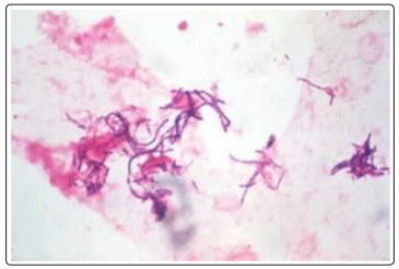
Figure 4: Erysipelothrix rhusiopathiae Gram stain [44].
Culture of E. rhusiopathiae from tissue specimens is relatively simple and requires only basic laboratory equipment and culture media such as tryptose or meat infusion media with or without blood or serum added. Care should be taken to avoid accidental skin infection, as the organism is pathogenic for humans. Selective culture methods for isolation of the organism from contaminate d specimens are described elsewhere [46].
The use of antiserum for treatment of suckling pigs is a fairly common practice. Initiation of a vaccination program in previously unvaccinated herds where outbreaks occur is strongly 15 recommended. For maximum effectiveness the serum must be given early in the course of the disease. The recommended therapeutic dose, given S/C, varies from to 5 to 10 mL for pigs weighing less than 50 pounds (23 kg). 20-40 mL for pigs weighing more than 100 pounds (45 kg). Penicillin alone is usually adequate when the strain has only mild virulence at Standard dose of 50 000 IU/kg BW of procaine penicillin I/M for 3 days [48].
Chronic cases do not respond well to treatment because of structural damage that occurs in joints and inaccessibility of the organism in the endocardial lesions. So there is no practical treatment for chronic SE. Experimentally, the administration of anti-inflammatory agents has provided some alleviation of the effects of chronic arthritis, and they may be used in treatment of especially valuable individual animals [49]. Fever associated with acute infections can be managed by administration of NSAIDs such as flunixin meglumine or by delivery of aspirin in the water. Erysipelas antiserum is described as an effective adjunct to antibiotic therapy in treating acute outbreaks but is not commonly available. Treatment of chronic infections is usually ineffective and not cost effective [50].Eradication of erysipelas on individual piggeries is considered impractical due to a large number of ?carrier? pigs and other ?carrier? species including birds and rodents. General hygienic precautions should be adopted. Clinically affected animals should be disposed of quickly and all introductions should be isolated and examined for signs of arthritis and endocarditis. All animals dying of the disease should be properly incinerated to avoid contamination of the environment. A combination of regular vaccination, good sanitation, the elimination of carriers with skin and 16 joint lesions, and appropriate quarantine measures for purchased stock usually will aid control of Swine Erysipelas [52, 53].
Vaccination against E. rhusiopathiae is very effective in controlling disease outbreaks on swine farms and should be encouraged. Vaccination using killed bacterins or, attenuated vaccines prepared by serial passage or strains of low virulence for pigs. The formalin-killed, aluminum-hydroxide-adsorbed bacterin confers an immunity that, in most instances, protects growing pigs from acute disease until they reach market age. An oral vaccine of low virulence is also used. Generally for prevention of SE all gilts and young boars should be vaccinated twice 2- 4 weeks apart (according to manufacturer?s instructions) before entering the breeding herd. Sows should be vaccinated 3-4 weeks prior to farrowing. Boars should be vaccinated every 6 months. In high-risk situations, vaccination of young stock from 6 weeks of age (either with a single dose or, if necessary, a 2 dose course) can be applied [54]. In addition to vaccination, attention to sanitation and hygiene and elimination of pigs with clinical signs suggestive of erysipelas infection represent other viable methods that may help control the disease on swine farms [55, 56].