Author(s): Le Phuc
A 30- year- old male has sustained open fracture of left leg and undergone 4 surgeries for four years (1. debridement and external fixator, 2. removal of external fixator, 3. vascularized contrafibular autograft plus change new plate/screws of ipsifibula, 4. flap surgery to cover the wound). At the time of examination: unability to walk, abnormal movements of leg, tibial bone loss 8 cm, especially the soft tissue of almost total anterior leg was so bad: no more subcutaneous tissue, skin very next to bone decouraging any attempt anterior approach. Diagnosis: pseudarthrosis of left leg with tibial large bone loss in the status of seriously damaged soft tissue of anterior leg surface. Technique operation chosen: Posterolateral. Appoach, Tibiofibular Fusion with massive autocancellous bone graft harvested from posterior iliac pelvis. Casting 4 months (from midthigh to metatarsal head). Postoperative walking with two crutches, Full Weight Bearing allowed after 14 weeks. Removal of cast after 16 weeks. Full knee flexion obtained 8 weeks after cast removal. Review at tenth month: normal walk, slight running, complete knee Range of Motion, 5. degree loss of dorsiflexion of ankle. X-rays: good fusion proximal and distal tibiofibular spaces and partly fusion the tibio-tibial gap. By this case,we can resumably conclude Tibiofibular Fusion through Posterolateral Approach should be strongly indicated for the leg nonunion in which the soft tissue of anterior surface is not favorable for nearby approach.
Nonunion of the tibia is a common complication of complex fracture of the leg. In general, it account for 5-20% of tibial fractures depend on severity extent of soft tissue and/or bone injuries caused by initial lesions and/or subsequent surgeries.
After Weber & Cech, there are 7 categories of nonunions: elephant foot (C1), horse’s foot (C2), oligotrophic (C3), dystrophic (torsional wedge with butterfly) (C4), necrotic (comminuted or avascular fragment) (C5), bone loss (C6), atrophic (C7). Conservative treatment has a modest role in nonunion; operative treatment by several techniques have proven effective in majority of cases. The main steps in surgery include debridement and refreshing the fractures, stripping the periosteum and bone grafting.
If the soft tisse of anterior leg is so destructive, and the alignment of tibia and fibula were in the range of acceptability, Tibiofibular Fusion by Posterolateral Approach should be indicated as the best treatment. This surgical technique was applied successfully in
USA by Freeland, Hanson, Jones and in France and francophone countries by Evrard, Vidal, and Moyikoua [1-3]. Tibial nonunion are exposed as large as possible, debridement, fractures freshened. Cancellous bone harvested from posterior iliac are placed and filled in the proximal and distal tibiofibular spaces and the gap between tibial fragments. Immobilisation (by cast or external fixation) is maintained for an appropriate length [4-9].
This paper, presents one case of large tibial bone loss treated by above-mentioned method.
Patient is a male 30 years old, had an complex fracture of left leg four years ago. He experienced 4 surgeries 1) Debridement and stablized the tibia by external fixator. The fibula was fixed by plate and 6 screws. Figure 1. 2) Removal of external fixator 3) Vascularized contrafibular autograft and changed the plate & screws of ipsifibula (new plate with 8 screws). Figure 2. 4) Flap surgery to cover the wound. X-rays showed contrafibular bone graft well reabsorbed, not rigid enough for weight bearing. Diagnosis: nonunion with large bone loss of left leg. One challenge: soft tissue of anterior leg was so bad, no longer subcutaneous tissue, skin very near bone which decouraged any approach nearby Figure 3.

Figure 1: X-ray After Removing External Fixator. First Operation: Debridement of Open Fracture of Left Leg. Fibula was Stablized by Plate & 6 Screws. Tibial Large Bone Loss 8 cm

Figure 2: Vascularized Bone Graft used the Contrafibula. Change the Plate & Screws of Ipsifibula by New Plate and 8 Screws. 3 Months after, the Fibula Bone Graft Seemed Integrated Well With Host Tibia
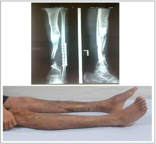
Figure 3: 6 Months after Surgery, The Contrafibular Bone Graft well Reabsorded, Not Rigid Enough for Weight Bearing. Soft Tissue of Anterior Leg was so Bad: Skin very near bone, No more Subcutaneous Tissue. Approach from nearby Anterior was Strongly Contraindicated.
Patient was treated “Tibio-Fibular Fusion through Posterolateral Approach”. Prone position. Tourniquet at groin. Skin incision 8cm longitudinal on the posterolateral of leg, 1-2 cm posterior to the fibula. Good exposure of the gap between tibial, and the tibiofibular proximal and distal spaces. Posterior tibial artery and veins were not exposed but well protected in the muscular mass. Chipbones of autologous cortical cancellous bone grafts harvested from posterior iliac wing were placed and filled in tibiofibular spaces and between two tibial fragments as well. Figure 4. Casting from midthigh to head of metatarsals for 4 months. Figure 5. Walking with 2 or 1 crutch and Partial Weight Bearing permitted during 3 first months. Full Weight Bearing allowed from the end of third month. Removal of cast at the end of fourth month.

Figure 4: Operative Technique, Tibiofibular Fusion by Posterolateral Approach

Figure 5: X-ray After Tibiofibular Fusion Surgery
After removal of cast: able walking with 1 or 2 crutches. Knee ROM 0/0/50. X-ray: bone developed, partly fused tibiofibular spaces and the gap between tibial fragments. Figure 6.
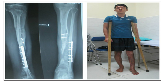
Figure 6: Patient After Removal of Cast, Able Walk With 2 or 1 crutch. Knee ROM: 0/0/50.X-ray: Bone Developed and Fused Tibiofibular Spaces and The Gap Between Tibials
Reexamination at 10th month: walking almost normal, slight running. ROM: 0/0/1200. X-rays: bone developed & fused well tibiofibular spaces. Good bone healing the gap between tibial fragments. Figure 7.
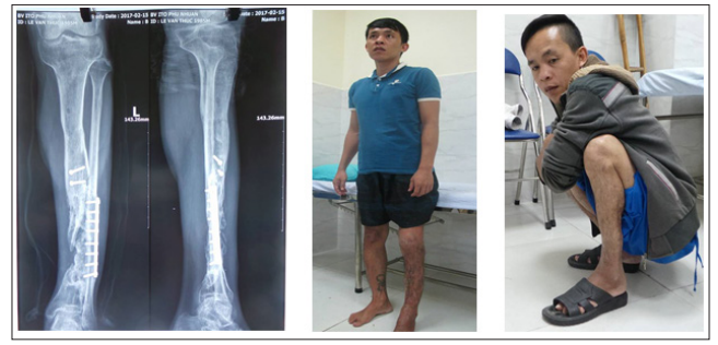
Figure 7: 10th Month Reexamination: Walking Almost Normal, Slight Running. ROM 0/0/1200.X-Rays: Bone Developed & Fused Well Tibiofibular Spaces, And the Gap Between Tibials As Well
Large bone loss of tibia,if soft tissue of anterior leg in good status, the treatment is standadized: massive bone graft (autograft or allograft or plus bone substitute) after debridement, removal of strange materials, freshening the fragments until bloody, stripping the periosteum from bone to stimulate bone healing, then immobilisation by cast or internal or external fixation. In case that soft tissue of anterior leg is seriously destructive, the management must be different: not routine anterior approach, not routine site to harvest bone for grafting.
For this special situation, the approach accepted by majority of orthopaedic surgeons is posterolateral which avoid the scar, preserve well the vulnerable anterior leg, access not so difficultly the nonunion, expose and freshen tibial fragments, and place the bone graft. Posterolateral approach for tibiofibular fusion first time described by Harmon in 1945, since then it’s proven more and more useful [4]. Tibiofibular fusion is one way to achieve bone healing of the leg in which the fibula healed well or not fractured, rigid enough for the weight bearing. There are some techniques to fuse tibiofibular as Merle D’Augbine’s, or Campanacci’s or Duparc’s [8]. For this patient, chipbones were used, filled in the gap between two tibial fragments and tibiofibular spaces. No hardware or a big piece of bone graft were utilized to stablize the fracture [6].
By this one case report, the Tibofibular Fusion by Posterolateral Approach would be strongly recommended for leg nonunion (with bone loss or not) in which anterior soft tissue were so damaged, but tibial and fibular alignment were in the range of acceptability.
No funding or grant support was received for this work.
No financial disclosures